The MU School of Medicine’s Craniofacial Research Center is committed to supporting state-of-the-art clinical and translational research to advance the understanding of the factors influencing craniofacial growth, development and form.
Craniofacial anomalies are among the most common congenital defects in humans, affecting as many as 1 in 700 births. Even minor conditions can have a dramatic impact on the psychological well-being of individuals throughout their lifetime. The Craniofacial Research Center currently includes a team of researchers and clinicians making novel discoveries in multiple aspects of craniofacial biology and treatment.
Research projects underway in the center include the Craniofacial Growth Consortium Study (CGCS) led by director Richard Sherwood, PhD. This study combines head radiographs (aka lateral cephalograms) from the historical archives of eight of the largest growth studies in North America. These studies began as early as 1910 collecting longitudinal data on growth and development from birth to adulthood. The CGCS now contains long-term growth data on 2,100 individuals. Using newly developed statistical approaches, the CGCS is creating growth standards and models that will provide clinicians with new prognostic tools for evaluation of current craniofacial morphology and prediction of patient growth potential and craniofacial milestones. These tools will aid clinicians in making evidence-based decisions that will optimize treatment timing.
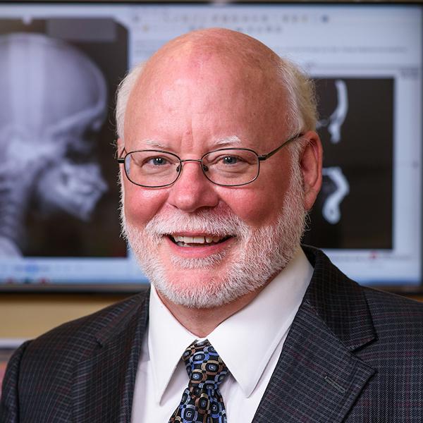
Richard Sherwood, PhD
Science has a difficult time explaining unique events. Fortunately, in the natural world, unique events are rare. Human biologists are faced with defining, describing and trying to explain a set of unique characteristics on a regular basis. Human biology is characterized by a suite of unique traits involving our locomotion, life history, reproductive physiology and cognitive ability. The human craniofacial complex is no different, with the unique combination of flexed cranial base, orthognathic face, and large, globular neurocranium. The usual strategy of the comparative biologist is to collect data on homologous structures and behaviors from — preferably closely related — animals and use this to test hypotheses regarding the animal of interest. In this work, traits may be considered either independently or as part of a morphological, functional or developmental complex. Anthropologists and human biologists again are hindered as no animal, including our closest living relatives, share any of our most interesting traits, even when taken as independent entities, of primary interest.
While this may seem to place the comparative anatomist in futile pursuit of an unobtainable goal, this is not the case. It merely necessitates the examination, and understanding, of every potential clue to the puzzle. In order to obtain an understanding, the examination must take place on many levels. As an anatomist, much of Dr. Sherwood’s work involves identification of traits and characterizing their variability in either quantitative or qualitative terms. As a comparative anatomist, he is then required to examine similar traits in additional populations, characterize the variability within these populations, and note the similarities or differences within the initial population. Then, where possible, Dr. Sherwood extends this approach into the fossil record to make inferences about the historical processes responsible for the observable morphology.
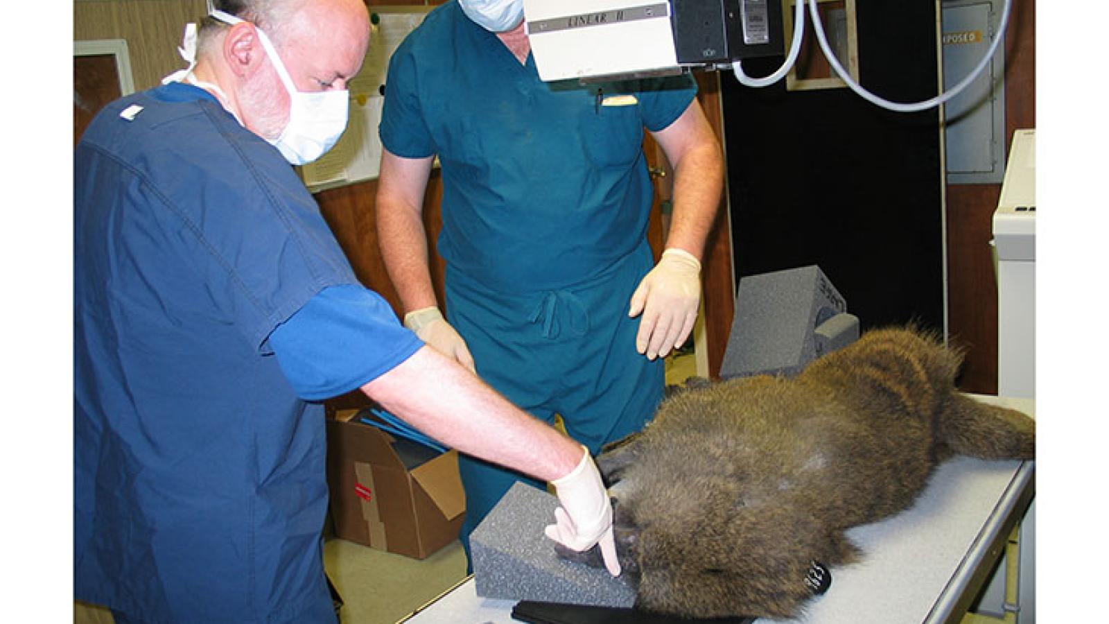
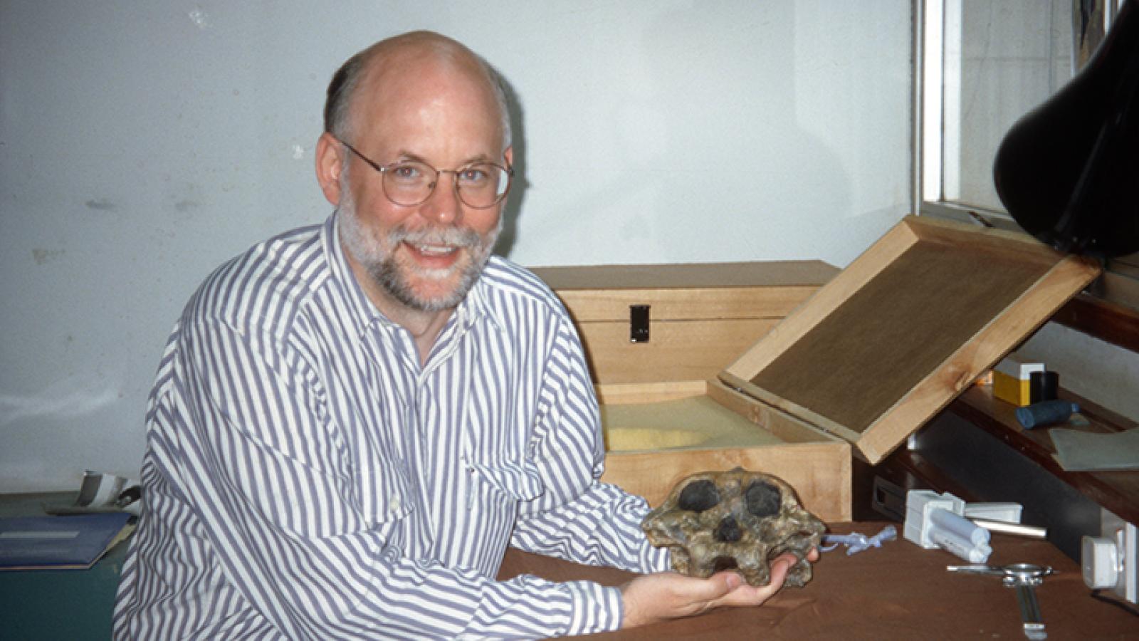
His career has focused on understanding the craniofacial biology of humans and non-human primates. In this work he has examined the morphology of numerous fossil and modern skulls both externally and internally (with the help of radiographic techniques such as CT imagery) from around the world. Dr. Sherwood has used analytical approaches, from simple univariate statistics to complex geometric shape analyses, with current analyses directed at using state-of-the art quantitative genetic approaches to detail the genetic architecture of the craniofacial complex. In this work his goal has been to understand the forces, genetic and non-genetic, influencing variation of the craniofacial complex across primates.
Dr. Sherwood’s current work explores craniofacial growth with novel approaches to longitudinal modeling. This project has roots dating back to 2007, when he became a founding curator of the American Association of Orthodontists Foundation (AAOF) Legacy Collection, a virtual collection of cranial radiographs from eight of the largest growth studies in North American (https://www.aaoflegacycollection.org/). He was able to leverage the Legacy collection to create the Craniofacial Growth Consortium Study. This work, funded by the National Institute of Dental and Craniofacial Research, is now the largest longitudinal study of craniofacial growth having assessed more than 17,000 radiographs. Please explore this site more to find out about the research projects currently underway.
Read more about Dr. Sherwood: Dr. Sherwood one of four MU faculty honored as 2024 AAAS fellows
Kevin M. Middleton, PhD
Kevin Middleton, PhD leads the biostatistical analyses for the Craniofacial Research Center. During his career, Dr. Middleton has developed innovative statistical and analytical approaches to challenging questions. Much of his research has been focused on the integrative biology and evolution of vertebrate tissues in model and non-model systems. This work spans biostatistics, computationally intensive methods, comparative anatomy, functional morphology, and biomechanics.
With extensive experience in data and statistical analyses, including both frequentist and Bayesian methods, mixed- and longitudinal-models, multivariate methods, nonlinear models, geometric morphometrics, and machine learning Dr. Middleton directs the statistical analysis for all aspects of the craniofacial projects covered at the center, including the non-linear growth models successfully used to estimate craniofacial growth.
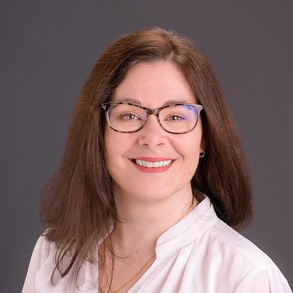
Dana L. Duren, PhD
Dana Duren, PhD is a leader in bone and joint health across the lifespan. As an expert in somatic maturation, she has expertise in a wide variety of methods for assessing skeletal and dental maturation using medical imaging. Within the Craniofacial Research Center, Dr. Duren is spearheading the efforts to incorporate maturation status into craniofacial growth models. When growth is assumed to track chronological age we do a disservice to patients who have unique lived experiences and backgrounds. Alternatively, tracking individuals according to the more biologically-meaningful skeletal age can significantly improve the evaluation of growth status and growth potential at an individualized level. Current work in the center will improve individualized models predicting the magnitude and timing of facial growth by incorporating objective measures of skeletal maturity of the individual better approximating the timing of somatic growth than can be achieved using chronological age.
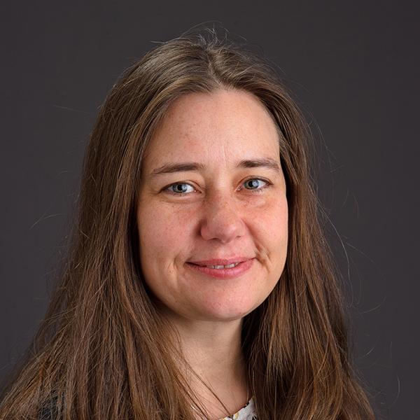
Kristina Aldridge, PhD
Kristina Aldridge, PhD, uses her background in neurobiology and craniofacial anatomy to examine the interactions between brain and bone during development. Dr. Aldridge’s work includes research on craniosynostosis, a disorder of premature suture closure altering head shape in young children. This work suggests a substantial degree of independence and interdependence in brain and skull development not previously appreciated. This suggests that the neuroanatomical basis for mild cognitive deficits observed in children with craniosynostosis are likely localized rather than global changes. These unprecedented studies of the brain in a craniofacial disorder have led to a paradigm shift from viewing the brain as a passive structure in head growth. They have also led investigators around the country to examine the neuroanatomical basis for the mild cognitive deficits observed in children with relatively mild craniofacial disorders.