
As an undergraduate anthropology student at the University of Missouri, Dana Duren wondered why humans walk on two feet while their closest animal relatives get around on four. Duren’s mentor, Carol Ward, PhD, suggested she compare X-ray images of the thigh bones of humans and chimpanzees.
“If you look at an infant human femur and an infant chimp femur, they look very similar,” Duren said. “But an adult of each of those looks completely different.”
The radiographs showed that as humans grow, their femurs start angling in from the hips, positioning their feet under their center of gravity. Chimps’ femurs grow straight, giving them a stance too wide to walk comfortably on two legs.
The evidence was all there in black and white.
That project piqued Duren’s interest in what other human-development questions could be answered by examining bones. This interest led to her current position as the MU School of Medicine’s director of orthopaedic research and professor of orthopaedic surgery. Duren is now in the ninth year of a study funded by the National Institutes of Health that examines trends in skeletal maturity. It has yielded noteworthy results.
In September 2018, she published an attention-grabbing article in the journal Clinical Orthopaedics and Related Research that showed children are growing up faster than before.
“Our findings show there is a ‘new normal’ for timing when kids’ skeletons will reach full maturity,” Duren said.
Once again, the proof was in the X-rays, if you knew where to look.
The Truth is in There
After earning her PhD in biological anthropology at Kent State University, Duren began her career in 2001 at Wright State University’s School of Medicine. It was a good fit for her interests, because the school was managing a long-running human development project called the Fels Longitudinal Study.
Beginning in 1929, parents in the small Ohio town of Yellow Springs enrolled their children in the study. In the name of science, the participants regularly were measured, monitored and scanned as they grew into adulthood. They often enrolled their own children in the research project, which ended in 2018.
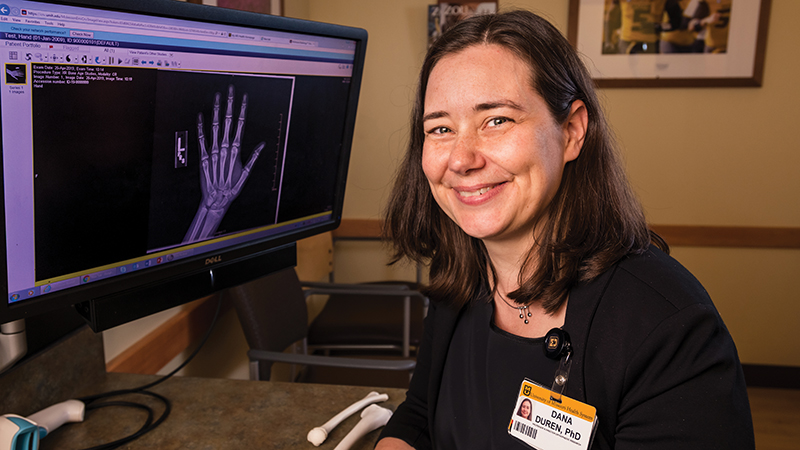
The Fels study created a rich vein of data for Duren to mine, first at Wright State, then at MU, starting in 2016. For the latest project, Duren and her team examined radiographs of the left hands and wrists of 1,292 children born between 1915 and 2006.
To completely analyze a hand-and-wrist X-ray using the established Fels Method for Skeletal Maturation requires a meticulous review of 98 features, but Duren and her team focused specifically on epiphyseal fusion.
Growth in long bones takes place in the cartilage plate near the end. When the end — the epiphysis — builds a bone bridge across the cartilage plate to the shaft of the bone, that is known as epiphyseal fusion. It signals the bone is done growing.
The research showed boys born in 1995 reached skeletal maturity in some bones about seven months faster than those born in 1935, while girls matured almost 10 months faster.
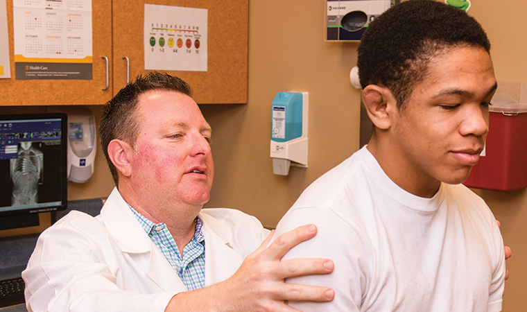
Duren’s study did not address the cause of the acceleration, and she doesn’t see earlier maturation as inherently good or bad — it’s just important information for clinicians. In fact, the research is already being applied at MU Health Care. Pediatric orthopaedic surgeon Daniel Hoernschemeyer, MD, collaborates with Duren’s team as he assesses when scoliosis patients need treatment, such as vertebral body tethering.
“When we consider VBT surgery for a child with scoliosis, there are concerns with overcorrection,” Hoernschemeyer said. “Understanding maturation and growth helps us with the timing of surgery to lower the risk of this issue.”
Just Getting Started
As an undergrad, Duren found a valuable mentor in Ward, who is currently a curator’s professor in the MU School of Medicine’s Department of Pathology and Anatomical Sciences. Now, Duren is the mentor.
One of her star pupils is PhD candidate Melanie Boeyer, who shares Duren’s enthusiasm for the subject manner.
“Halloween was always my favorite day,” Boeyer said. “I dressed up as a skeleton. I wore anything that had a skull on it. You know the plastic skeletons you put out in your yard to scare people? I had those in my room as decorations all year long.”
After reading one of Duren’s previous publications, Boeyer asked to join Duren’s Skeletal Morphology Laboratory at MU. For the latest study, Boeyer helped to analyze the data and served as first author.
“We assume we know a lot about how kids grow and develop, but we have a lot more to learn,” Boeyer said. “This is a pretty good way to assess what we know and what we don’t know. What we’re seeing is kids now are not the same as kids way back when.”
As helpful as the Fels study was in showing bone fusion changes over time, it has its limitations. Ninety-eight percent of the participants were white. Moving forward, Duren and her team want to further modernize skeletal maturity standards.
“We don’t want to be looking at an African-American boy today and be comparing him to a white boy from the 1930s. That just doesn’t make any sense,” Duren said. “My grant through NIH updates those methods. I’ve started collaborating with colleagues who have conducted a study of bone-mineral density in childhood that has groups of children from five different sites across the U.S. We have African-American, Asian-American, Hispanic and white — all born after 1990. With 8,000 films from those kids, we’re redoing this.”
When the project is finished, Duren’s team will deliver the most comprehensive method for skeletal-maturity assessments that is applicable to modern children of multiple backgrounds, which will help clinicians make the best decisions for their growing patients.